
A pharyngeal pouch is like a hernia at the top of your oesophagus. They usually grow slowly and it can be many years before patients realise they have problems. While some pouches do not cause any problems, some can cause difficulty swallowing, regurgitation of food and lead to chest infections.
In these cases Professor Winter would often advise monitoring your swallow. However when the pouch is causing problems then there are 2 main operations for treating a pharyngeal pouch:
- Dohlman’s Procedure (endoscopic stapling or laser technique)
- An open operation to close the pouch off
Normal pharyngeal function
The normal pharynx acts as a passage between the mouth and oesophagus. When one swallows food, it passes from the mouth to the pharynx and through to the upper oesophagus.
For food to enter the upper oesophagus, a muscular valve at the top of the oesophagus (known as a sphincter) must relax as the food approaches. The muscle called cricopharyngeus forms this valve or sphincter.
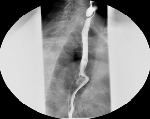
A pharyngeal pouch, also known as a Zenkers diverticulum is an outpouching of the pharynx, just above the cricopharyngeus muscle. The cause of this outpouching is due to either uncoordinated swallowing or impaired relaxation and spasm of the cricopharyngeus muscle. The cause of this may be due to chronic acid reflux.
The increased pressure within the pharynx, causes its wall to herniate through the point of least resistance (Killian’s dehiscence). The result is an outpouching of the posterior pharyngeal wall, just above the oesophagus.
Endoscopic Stapling/Laser
Endoscopic treatment is an accepted way of treating these pouches and usually requires one night in hospital. On the day of the operation you will meet with myself and the anaesthetist.
At the operation I will pass a rigid endoscope through the mouth to identify the pouch and inspect the lining with a camera. Depending on the pouch then either a laser will divide pouch wall or a stapling devise cut and staple the pouch wall with adjacent normal tissue so that food cannot get caught. This operation also involves cutting muscle band (sphincter) at the top of the oesophagus reducing the chance that this problem re-occurs.
Post-Operative Care
After the operation all patients are carefully monitored on the ward to make sure that they are recovering from the anaesthetic and also to identify any patient who is developing a complication of surgery, such as a leak in their oesophagus. Patients can usually start to drink fluids on the day of the operation and gradually build up to normal food. Patients are normally discharged the following day.
Complications – which are uncommon
Complications are rare but include damage to the teeth as the endoscope is passed. A hole developing in the stapling line allowing fluid or food to leak into the neck. If this occurs a feeding tube is usually passed to rest the area for a few days. Very occasionally an operation to open the neck and repair the hole is required. Finally in the long term the initial swallowing problem can recur caused by the pouch reforming. If this latter event does occur then the procedure can usually be repeated.
Open repair of the pouch
Certain pouches may be better treated with an open approach. This is usually done under general anaesthetic with a cut on the left side of the neck. Professor Winter will identify the pouch and surgically remove it. Professor Winter has performed this procedure regularly and is adept at discussing with you the merits of different approaches


